
What is BrainSaw?
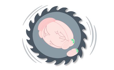
BrainSaw is an open-source, block-face serial section two-photon tomography (STPT) system for whole-brain imaging. It provides high-resolution, whole-organ volumetric imaging whilst avoiding complex sample preparation. In an STPT system, a multi-photon microscope is coupled to a vibratome and a three-axis stage. Fixed tissue is embedded in agarose and mounted in a water bath riding on the stage. The tissue surface is exposed by the vibratome and tile-scanned by the two-photon microscope. The STPT system alternates between slicing and imaging of the block face until all of the sample has been acquired.
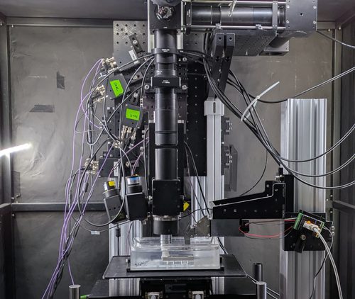
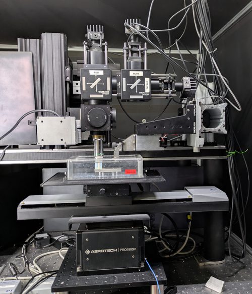
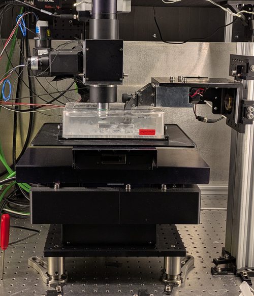
BrainSaw STPT was developed at the Sainsbury Wellcome Centre Advanced Microscopy Facility and is ideal for high-throughput imaging of whole mouse brains. Up to 6 mouse brains can be imaged overnight on a single microscope; the process is fully automated and highly reliable. The SWC's three BrainSaw systems (below) have imaged over 7,000 brains in the last decade, and the technology has been adopted by others and spread worldwide. BrainSaw has contributed to over 30 research papers and is routinely used for whole-brain imaging tasks such as mapping electrode tracks or brain-wide cell counting.
Our BakingTray acquisition software is modular and can run new custom STPT systems tailored to the needs of the host institute. It also serves as a platform for upgrading legacy TissueCyte 1000 systems from TissueVision, providing an open-source base upon which the system can be further modified. Existing two-photon microscopes can be converted to a BrainSaw for as little as 35,000 EUR. See our methods paper for more information and the news article on the main SWC website.
Ecosystem
BrainSaw: microscopy systems
We provide CAD models and build instructions for assembling a BrainSaw serial-section two-photon microscope. Systems can be built from scratch, or serial-sectioning functionality added to an existing microscope.

BakingTray: serial-section acquisition software
BakingTray is modular serial-section acquisition software for MATLAB. It accommodates diverse hardware configurations and acquires images via ScanImage. BakingTray can be used to build new custom STPT systems and upgrade legacy TissueCyte 1000 systems.
Docs
StitchIt: assembling tile scan data into stitched sections
StitchIt runs alongside BakingTray to pre-process data during acquisition, stream the last stitched section to a web page, and initiate stitching automatically when acquisition completes. It includes tools for downsampling and batch-processing image stacks, with operations highly parallelised for speed.
Docs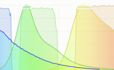
BrainSaw Channel Chooser
This interactive web tool helps BrainSaw users choose the best wavelength and excitation channels for their fluorophores.
Launch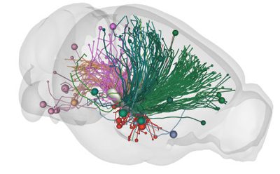
Analysis: BrainGlobe
Our favoured option for analysis of serial section data is the BrainGlobe suite. These Python-based tools are maintained by the SWC neuroinformatics team. BrainGlobe comprises several software packages to access, analyse, and visualise anatomical data.
brainglobe.infoVideo: BakingTray in action
This video shows acquisition of a single section on a BrainSaw serial-section two-photon microscope using BakingTray acquisition software. The video was acquired at the Sainsbury Wellcome Centre Advanced Microscopy Facility. We see acquisition of four mouse brains near the start of the process, at the cerebellum. More details are available on the YouTube page.