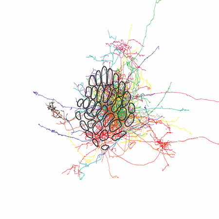
Single-axon tracing
Reconstructing the morphology of excitatory neurons in mouse barrel cortex, imaged with BrainSaw and registered to the Allen Common Coordinate Framework (CCF).

Researchers have used BrainSaw serial two-photon tomography to image thousands of brains, generating 3D datasets that can be automatically annotated and registered to a standard atlas for unbiased analyses. The system's major use cases are mapping electrode tracks, brain-wide cell counting, and tracing bulk axonal projections.
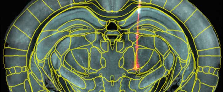
Electrodes can be coated with a dye that remains in the tissue after the electrode is withdrawn. Brains are imaged and the dye tracks can be traced and registered to a standard atlas. The brain areas through which each electrode passes can be determined automatically, and penetrations from multiple animals overlaid in a common space. This approach was applied at scale by the International Brain Laboratory (IBL), mapping 699 electrode tracks from 139 mice.
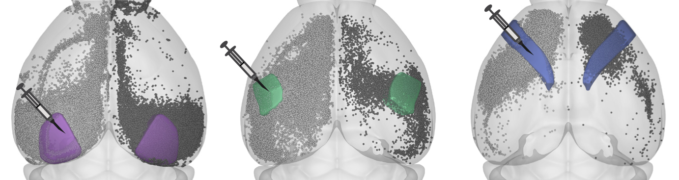
Techniques such as rabies tracing label individual neurons, revealing connectivity between brain areas. BrainSaw images these samples at high resolution, producing seamless 3D datasets. Automated tools, such as cellfinder, identify the locations of labelled neurons across the brain, allowing researchers to easily quantify cell numbers by brain area. If desired, the fluorescent protein can be localised to the nucleus, which makes automated cell detection easier.
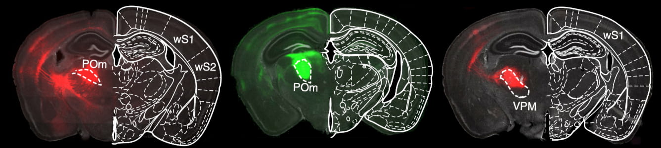
Cells can be induced to express fluorescent proteins with viral injections. The protein diffuses throughout each cell, filling somata, dendrites, and axons. Thousands of neurons are labelled by a large injection, and the regions to which the population projects can easily be determined by thresholding. The thresholded maps can be registered to a standard atlas for quantitative analysis. In this approach, single axons are not traced, so fine axial sampling is unnecessary.
Reconstructing the morphology of excitatory neurons in mouse barrel cortex, imaged with BrainSaw and registered to the Allen Common Coordinate Framework (CCF).
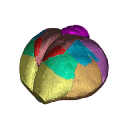
Generating a 3D template volume and brain atlas for the Eurasian blackcap.
Sirmpilatze et al., Current Biology, 2026
(using brainglobe-template-builder)
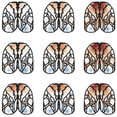
Using cortical blood vessels to project Allen CCF brain area borders onto widefield functional activity in dorsal cortex.
BrainSaw has also been used for methods development, screening AAV expression, validating whole-mount antibody staining, verifying virus injection sites, registering functional ultrasound data to the atlas, mapping lesions, and mapping the locations of implanted optical fibres.